How Does MRI Work?
MRI utilizes powerful magnetic fields and radio waves to generate detailed images of the body’s internal structures. To understand the process, we first need to look at the building blocks of our bodies. Most of the human body is composed of water molecules, which contain hydrogen atoms. At the core of each hydrogen atom is a proton, which behaves like a tiny magnet.
When a person lies inside the large, cylindrical MRI scanner, the powerful magnets within the scanner cause these protons to align in a particular direction, similar to how a compass needle aligns with the Earth’s magnetic field. Once the protons are aligned, short bursts of radio waves are emitted. These radio waves disrupt the alignment of the protons, causing them to absorb energy and change their orientation. When the radio waves are turned off, the protons gradually return to their original aligned state, releasing the absorbed energy in the form of radio signals.
The MRI scanner’s receivers pick up these signals, and a computer then processes the information to create detailed cross-sectional images of the body. Different tissues in the body, such as fat, muscle, and organs, have distinct proton densities and relaxation times (the time it takes for the protons to realign after the radio waves are turned off). This difference in proton behavior allows the MRI to distinguish between various tissues and create clear, detailed images.
The MRI Scanner: A Closer Look
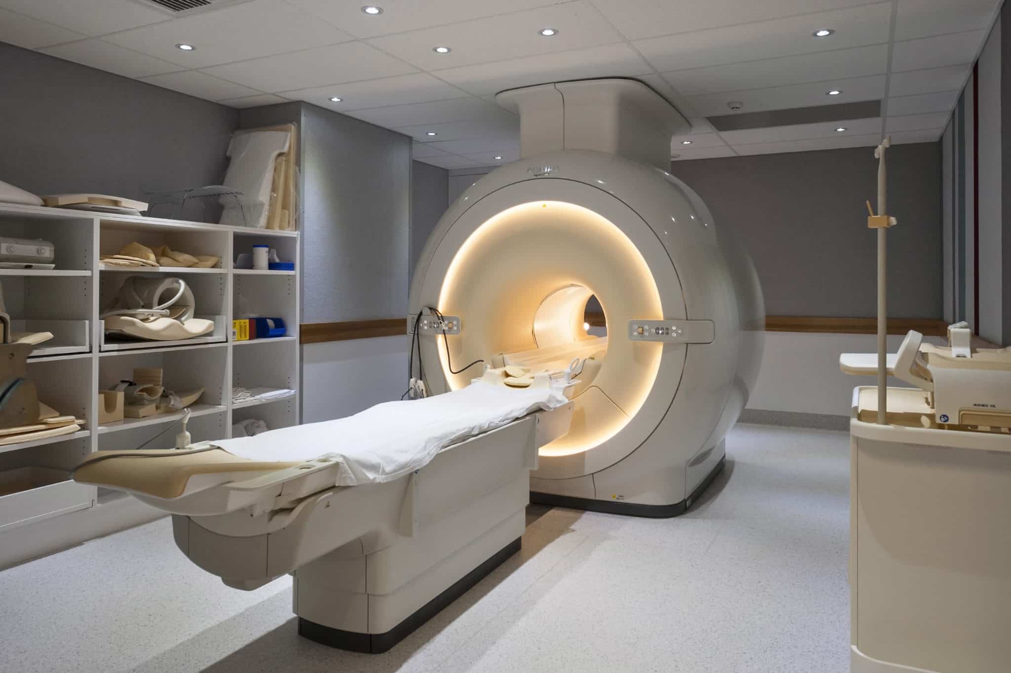
The MRI scanner itself is an imposing piece of equipment. It consists of a large, hollow cylinder with a narrow opening in the middle, where the patient lies. The scanner is equipped with multiple powerful magnets, typically superconducting magnets cooled by liquid helium to extremely low temperatures. These magnets create a magnetic field that is hundreds of times stronger than the Earth’s magnetic field.
There are also gradient coils within the scanner that can vary the magnetic field strength in different directions. This allows for the precise localization of the signals from the protons in the body, enabling the creation of detailed images. Additionally, radiofrequency coils are used to transmit the radio waves to the body and receive the signals back.
Applications of MRI
MRI has a wide range of applications across different medical specialties:
-
Diagnosing Neurological Conditions: MRI is extremely useful in detecting brain tumors, strokes, multiple sclerosis, and other neurological disorders. For example, it can clearly show the location and size of a brain tumor, helping doctors plan the best course of treatment. In the case of strokes, an MRI can identify the affected area of the brain and the extent of damage, which is crucial for timely intervention.
-
Evaluating Musculoskeletal Problems: It is commonly used to examine bones, joints, and soft tissues. Athletes with suspected ligament or tendon injuries often undergo MRI scans to determine the severity of the damage. For instance, an MRI can precisely diagnose a torn anterior cruciate ligament (ACL) in the knee, which is essential for deciding whether surgical repair is necessary.
-
Assessing Abdominal and Pelvic Organs: MRI can provide detailed images of organs such as the liver, kidneys, pancreas, and reproductive organs. It can detect liver diseases like cirrhosis, kidney tumors, and problems in the uterus or ovaries in women. For example, it can help in the early detection of ovarian cancer by visualizing any abnormal growths.
-
Cardiac Imaging: In the field of cardiology, MRI can be used to evaluate the structure and function of the heart. It can detect heart muscle damage, congenital heart defects, and problems with the heart valves. For instance, it can show how well the heart is pumping blood and identify any areas of weakened muscle.
The NHS and MRI Scans
In the United Kingdom, the National Health Service (NHS) offers MRI scans to patients when medically necessary. GPs or other healthcare professionals refer patients for MRI scans based on their symptoms, medical history, or as part of a diagnostic workup. The NHS has a network of hospitals and imaging centers equipped with MRI scanners. However, due to high demand, there may sometimes be waiting times for patients to receive their scans. For example, in some areas, the waiting list for an elective MRI scan for a non – urgent condition could be several weeks to a few months. But for urgent cases, such as suspected strokes or life – threatening tumors, patients are prioritized, and the scans are arranged as quickly as possible.
Private MRI Options
Private healthcare providers also offer MRI scans in the UK. Private MRI services often come with certain advantages. They may offer shorter waiting times, sometimes as little as a few days to a week, depending on the availability. Private clinics may also provide more convenient appointment times, including evenings and weekends, which can be beneficial for patients with busy schedules. Additionally, some private providers offer more specialized MRI services, such as high – field MRI (using stronger magnets for even more detailed images) or functional MRI (fMRI) for studying brain activity. However, the cost of private MRI scans can be a significant factor. A basic MRI scan in a private clinic can cost anywhere from £300 – £1000 or more, depending on the body part being scanned and the complexity of the scan.
Comparing MRI with Other Imaging Modalities
-
X – Ray: X – rays are useful for imaging bones and detecting fractures. However, they are not as effective in visualizing soft tissues like MRI. X – rays use ionizing radiation, which, although the risk is generally low with occasional use, is a concern with repeated exposure. In contrast, MRI does not use ionizing radiation, making it a safer option for many patients, especially those who may need multiple scans over time.
-
CT (Computed Tomography) Scan: CT scans are fast and can provide detailed images of the body’s internal structures. They are particularly good at detecting lung problems, such as tumors or blood clots. But like X – rays, CT scans use ionizing radiation. Also, in terms of soft – tissue resolution, MRI often provides more detailed images, especially for the brain, spinal cord, and joints. For example, when looking at a suspected brain tumor, an MRI can show more details about the tumor’s characteristics, such as its composition and relationship to surrounding tissues, compared to a CT scan.
https://about:blank/
|
Imaging Modality
|
Radiation Used
|
Soft – Tissue Resolution
|
Bone Imaging
|
Scan Time
|
Cost (Approximate in UK)
|
|
MRI
|
No
|
High
|
Not as detailed as X – rays for simple fractures
|
15 – 90 minutes
|
NHS: Free (at point of use); Private: £300 – £1000+
|
|
X – Ray
|
Yes
|
Low
|
Good for detecting fractures
|
Short (seconds)
|
NHS: Free; Private: Varies, around £50 – £200
|
|
CT Scan
|
Yes
|
Moderate
|
Good for complex bone and joint problems
|
Short (minutes)
|
NHS: Free; Private: £200 – £800
|
Data source: Compiled from NHS guidelines, private healthcare provider websites, and medical imaging research.
Preparation and Procedure for an MRI
Before an MRI scan, patients are usually given a set of instructions. Since the scanner uses strong magnetic fields, patients need to remove all metal objects from their bodies. This includes jewelry, watches, hairpins, piercings, and even some types of clothing with metal zippers or fasteners. Patients with metal implants, such as pacemakers, certain types of artificial joints, or metal plates in their bodies, may not be able to have an MRI scan, as the strong magnetic field can interfere with the implant or cause it to move.
On the day of the scan, patients are asked to lie still on a movable bed that slides into the scanner. Depending on the part of the body being scanned, they may be positioned head – first or feet – first. The scanner will make loud tapping noises during the scan, which is due to the switching on and off of the electric current in the scanner coils. To help with the noise, patients are provided with earplugs or headphones. The scan can last anywhere from 15 to 90 minutes, depending on the area being scanned and the number of images required.
QA about MRI
Q: Is an MRI scan painful?
A: No, an MRI scan is a painless procedure. However, some patients may find it uncomfortable due to the need to lie still for an extended period, especially if they have a condition that causes pain when remaining in one position. Also, the loud noises during the scan can be a bit disconcerting, but the provided earplugs or headphones help mitigate this.
Q: Can I eat or drink before an MRI?
A: In most cases, you can eat, drink, and take your regular medications as usual before an MRI. However, if the scan is of your abdomen or pelvis, you may be asked to fast for a few hours before the scan. Your doctor or the imaging center will provide specific instructions based on the type of scan you are having.
Q: Are there any risks associated with MRI?
A: MRI is generally considered a very safe procedure as it does not use ionizing radiation. However, there are some contraindications. People with certain metal implants, as mentioned earlier, may not be able to have an MRI. Additionally, in rare cases, some patients may experience a tingling sensation or feel hot during the scan. For scans that use a contrast agent (a substance injected to enhance the visibility of certain tissues), there is a small risk of allergic reactions or side effects such as nausea, headache, or a skin rash.


